A Closer Look at COVID Lung: Insights from COVID CT Scan
With the emergence of COVID-19 pneumonia as a global health issue, we take a look into how CT imaging has helped in diagnosing patients with the infectious disease.

COVID-19 pneumonia has emerged as a critical health issue globally, necessitating a comprehensive understanding of its manifestations and implications. Among the various diagnostic tools, CT imaging has gained prominence for its ability to reveal detailed images of lung pathology.
Understanding COVID-19 Pneumonia
COVID-19 pneumonia occurs as a result of infection by the SARS-CoV-2 virus, which primarily targets the respiratory system. This condition is characterized by inflammation in the lungs, leading to severe respiratory symptoms and complications. Understanding the nature of COVID-19 pneumonia is crucial for effective diagnosis and treatment.
The Role of CT Imaging in Diagnosing Lung Infections
CT imaging plays a vital role in diagnosing lung infections, offering high-resolution images that can reveal subtle changes in lung architecture. This imaging modality is particularly beneficial in assessing the severity and extent of COVID-19 pneumonia.
The high sensitivity of CT scans enables the detection of early pathological changes, which are critical for effective diagnosis and timely intervention in respiratory diseases.
In the context of COVID-19, CT imaging can showcase multifocal areas of involvement, aiding in determining the stage of the disease. The ability to reconstruct images in 3D, such as through the 3DICOM software, enhances the physician’s understanding of the spatial relationships between various structures within the lungs, providing a more comprehensive view of the infection’s impact.
Empowering Precision in
Modern Healthcare!
Modern Healthcare!
3DICOM MD, improves clarity and supports informed decision-making, simplifying complex medical information for both you and your patients.

Common Features of COVID 19 Pneumonia CT Imaging
Common findings in COVID-19 pneumonia include bilateral ground-glass opacities, consolidative areas, and interlobular septal thickening.
Ground-glass opacities can indicate the early stages of infection and pulmonary inflammation. As the disease progresses, these opacities may evolve into more pronounced consolidations, reflecting the ongoing inflammatory response and potential for secondary bacterial infections.
Symptomatic patients may also exhibit parenchymal abnormalities that evolve over time. It is important for health professionals to be aware of the evolving nature of these findings as they may correlate with the progression of COVID-related pneumonia.
For instance, the presence of crazy paving patterns—characterized by interspersed ground-glass opacities and reticular patterns—can suggest a more severe inflammatory process and may indicate a higher risk of respiratory complications.
Understanding the timeline of these imaging changes can be crucial for monitoring disease progression and guiding therapeutic interventions.
Interpreting CT Imaging Results
Accurate interpretation of CT findings is critical, as not every abnormality points directly to COVID-19 pneumonia. Radiologists must differentiate COVID-19-related changes from other types of pneumonia or lung conditions.
Moreover, the context of clinical presentation must guide interpretation, as certain features may overlap with non-COVID pulmonary pathologies.
For example, findings such as bronchial wall thickening or pleural effusions may suggest alternative diagnoses, including bacterial pneumonia or heart failure, necessitating a careful and nuanced approach to imaging interpretation.
Consultation between radiologists and treating physicians enhances diagnostic accuracy, allowing for the establishment of a diagnosis based on a combination of clinical symptoms and imaging findings. This collaborative approach aids in formulating treatment strategies tailored to individual patient needs.
As research continues to evolve, the role of advanced imaging technologies and AI in analyzing CT scans may also contribute to more rapid and precise interpretations, ultimately improving patient outcomes.
The Need for Further Research
Despite the advancements in imaging techniques, further research is required to enhance the understanding of COVID-19 pneumonia and its manifestations on CT scans.
Continued investigation into optimal imaging protocols and the development of standardized reporting systems can help physicians in making accurate diagnoses.
Want to explore COVID pneumonia on CT scan in 3D?
Find a free DICOM download of an infected lung CT in our DICOM Library, which can be explored in immersive 3D in the 3DICOM Viewer.
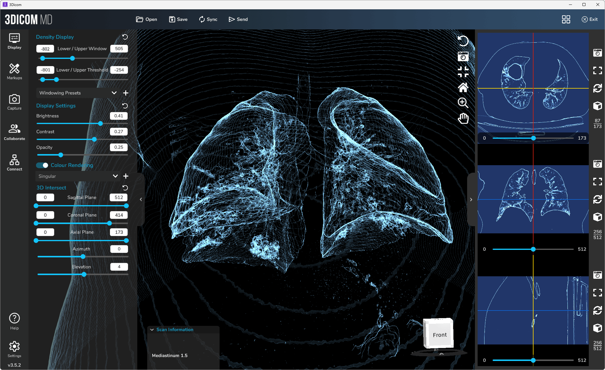
Read more about the 3DICOM MD Viewer featured in the screenshot above.
Explanation of Key Terms
Explanation of Key Terms
Modality
In medical imaging, the term modality specifically refers to the different imaging techniques used to create images of the body E.g. MRI, CT scans, and ultrasound. Each offers unique insights into the body’s internal structures and functions.
Ground-glass opacities (GGO)
Ground-glass opacities are findings on CT scans that indicate areas of increased lung opacity with preserved bronchial and vascular markings. The appearance is similar to frosted glass, suggesting partial filling of air spaces in the lungs or interstitial thickening. GGOs are non-specific and can be seen in various pulmonary conditions, including infectious and inflammatory processes, such as viral pneumonia or interstitial lung disease.
Interlobular Septal Thickening
Interlobular septal thickening refers to the enhancement of the lines in lung imaging that demarcate the boundaries between secondary pulmonary lobules. It is visible in high-resolution CT scans and indicates fluid, cellular infiltration, or fibrosis within the interlobular septa. This finding is common in conditions involving pulmonary edema, lymphatic spread of carcinoma, and various interstitial lung diseases, helping to differentiate among these pathologies based on their distinct patterns.
Parenchymal Abnormalities
Parenchymal abnormalities refer to changes or damage to the lung tissue itself, which can be seen on imaging studies like CT scans. These abnormalities may include a range of pathological changes, such as consolidation, fibrosis, nodules, or cavitations. Such changes are indicative of underlying disease processes affecting the lung parenchyma, including infections, inflammatory disorders, or neoplastic conditions.
Crazy Paving Patterns
Crazy paving patterns describe the appearance on lung imaging, particularly on high-resolution CT scans, where there is a combination of ground-glass opacities with superimposed interlobular septal thickening and intralobular lines. This pattern resembles irregularly paved stones, hence the name. Initially described in patients with pulmonary alveolar proteinosis, this finding is now associated with a variety of acute and chronic lung infections, inflammatory conditions, and diffuse lung diseases, reflecting alveolar filling with additional interstitial
Bronchial Wall Thickening
Bronchial wall thickening is a radiologic finding often observed on CT scans of the chest, where the walls of the bronchi appear abnormally thick. This can be due to inflammation, infection, or chronic irritative processes. It is commonly seen in conditions such as chronic bronchitis, bronchiectasis, and asthma. The thickening can be diffuse or localized, and its presence helps in diagnosing and assessing the severity of the underlying respiratory condition.
Pleural Effusions
Pleural effusions refer to the accumulation of excess fluid between the layers of the pleura, the thin membrane that lines the outside of the lungs and the inside of the chest cavity. This condition can cause chest pain and difficulty breathing and is visible on imaging studies such as ultrasound, X-rays, or CT scans. Pleural effusions can result from a variety of causes, including heart failure, pneumonia, liver disease, or malignancies, and identifying the underlying cause is essential for appropriate management.
Summary of Key Points
Summary of Key Points
COVID-19 Pneumonia Overview:
- Caused by the SARS-CoV-2 virus targeting the respiratory system.
- Characterized by lung inflammation leading to severe respiratory symptoms and complications.
CT Imaging‘s Role in COVID Diagnosis and Treatment Planning:
- Essential for diagnosing lung infections, particularly COVID-19 pneumonia.
- Provides high-resolution images that detect early pathological changes and assess disease severity.
- Showcases multifocal areas of lung involvement and can be used to reconstruct 3D images for a better understanding of lung architecture.
Common COVID 19 CT Scan Features:
- Bilateral ground-glass opacities and consolidative areas are typical in early infection stages.
- Interlobular septal thickening and crazy paving patterns indicate more severe disease and higher complication risks.
Interpreting CT Results:
- Critical to differentiate COVID-19 related changes from other lung conditions.
- Requires correlating CT findings with clinical presentation for accurate diagnosis.
- Collaboration between radiologists and clinicians is vital for effective diagnosis and treatment planning.
Advancements and Future Research:
- AI’s role in analyzing CT scans promises quicker, more accurate interpretations.
- Ongoing research is needed to improve imaging techniques, optimize imaging protocols, and develop standardized reporting systems.