
Experience empowering, portable, secure access to medical imaging— Anytime, Anywhere with 3DICOM.
★★★★★
Trusted by over 5,500+ medical practitioners, academics, and patients.
Your single unifying medical imaging software solution
Connecting people, devices, and systems for better health outcomes
Interactive 3D Visualization
Easily and securely convert 2D DICOM images from CT, PET, and MRI scans into interactive 3D models—quickly, on any device for better visibility and understanding.


Cross-Platform Availability
Conveniently access and securely store and share your medical images across platforms and devices with iOS, Android, Mac, and Windows compatibility, and benefit from immersive 3D viewing on XR devices.


Collaborative Tools and AI
Leverage AI and collaborative tools designed for effective communication and Telehealth1, treatment planning, and education. Streamline workflows with built-in screen recording, comprehensive annotation, measurement, and markup tools, and access a selection of AI models available in the cloud.
Convenient File Sharing and Interoperability
From PACS Integration to the ability to securely share medical imaging between devices, across platforms and between facilities, 3DICOM sets a new standard for interoperable medical imaging and cross-facility sharing in healthcare.
Move into a new era of convenience, and see for yourself!


What can 3DICOM do for you?
Improve Patient Outcomes
Benefit from better access to medical imaging, enhanced diagnosis and treatment planning, and improved patient communication.
As a patient, save yourself unnecessary scans and delays, be more actively engaged and informed about your healthcare, and accelerate your pathway to effective treatment.
Experience Effortless Access
Access and interact with medical images anytime, anywhere, on any device.
With 3DICOM’s seamless access and sharing, medical imaging is always at your fingertips—wherever you are, whenever you need it!
Transform Medical Understanding
Transforms complex 2D medical imagery into versatile, interactive 3D visualizations with the 3DICOM software for better visibility into the body.
This enhanced view helps improve the understanding of patient-specific pathologies and treatment options.
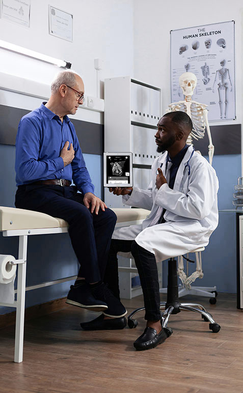
Enhance Collaboration and Education
Enabling remote consultations and collaborative planning, 3DICOM also serves as an educational tool for medical students and professionals.
It enhances communication and coordination, allowing teams to tackle complex medical challenges more efficiently and expedite innovative solutions.
AI Integration
Streamline education and workflows with AI
For patients: Feel empowered through enhanced understanding of your body
With AI integrations such as Total Segmentator and ChatGPT 4.0, which work alongside 3DICOM’s 3D visualization to help identify, label and educate you on anatomy, you can learn about your body and condition more easily, enabling you to make more informed medical decisions.
For medical professionals: Streamline workflows
As doctors and medical professionals, artificial intelligence models available with 3DICOM MD will help you get the most out of any medical imaging and streamline clinical workflow.
For academics: Accelerate research and development
If you are an academic or a researcher, the combination of 3DICOM EDU and advanced AI models improves workflows and accelerates the development of innovative patient-specific solutions.


Convenience
Move into an era of convenience
For patients: Access to your medical imaging is now in your pocket
With the ability to store and share your medical imaging directly from your mobile phone or online, your medical images can be viewed anywhere, anytime.
For medical professionals: Interoperability means you can say no to duplicate scans
With PACS integration and convenient accessibility and sharing at your fingertips, waiting on patient imaging and duplicate scans can become a thing of the past, allowing for better continuity of care for your patients.
#DitchtheDisk and say goodbye to CDs
By enabling medical images to be stored, accessed and shared instantly from any location, you can remove outdated CDs from the equation – and with them, remove the unnecessary costs, delays, and inefficiencies they create.
Resources to educate and inspire!
Explore our FREE library of open-source DICOM files.
All files are anonymized and ready to use for research and educational purposes.
-
-
R&D has moved online
3DICOM R&D is now entirely cloud-based As of September 1, 2025, 3DICOM R&D has officially transitioned to a fully cloud-based…

-
auto_storiesRadiology 101
Top 5 DICOM Viewers for Mac in 2024
Want to know the best DICOM Viewers to use on your Mac? Here is a list of some outstanding DICOM…

Hear what our customers have to say
“I now have one place where I can digitally store all of my information. 3DICOM has finally given me control over my medical imaging.”

Ashley C, Miami Florida
Patient
“The combination of the 3DICOM software and the SRD screen can make a difference to patient education, and if they are more involved, the outcome will be better.”

Alex S, Sion Switzerland
Neurosurgeon
“Thanks to 3DICOM’s new web portal, all of our medical images are kept online. All in one place and easy to find. It is more than convenience, it is peace of mind.”

Warwick R, Miami Florida
US Airforce Veteran
Learn more about our story
Read about 3DICOM’s beginnings and mission on our About page.

Unlock the full value of your medical imaging
Sign up today or try the FREE demo to experience the difference for yourself.
- Real-time collaborative Telehealth tools are only available in the FDA-Cleared 3DICOM MD Desktop Application. ↩︎