
Products
3DICOM EDU
Transform medical education with unlimited interactive and immersive medical cases and accelerate research and development through advanced collaborative and prototyping tools.
Designed for teachers, institutions, academics, students, and researchers.
ADD ANOTHER DIMENSION
to medical education!
Replace expensive cadaveric studies with immersive unlimited 3D anatomical and pathological cases for a more accessible and engaging experience for medical students.
COLLABORATE
for faster innovation!
Seamlessly share 3D anatomical models—complete with markups, overlays, clinical notes, and design files—to enhance communication and bridge the gap within multidiscipline teams.
PUSH THE BOUNDARIES
of medical research!
Accelerate the development of innovative patient-specific solutions by combining 3DICOM EDU’s automatic segmentation and rapid prototyping tools.


What’s Included

3DICOM EDU is trusted by the following partners:
VIDEO
Demonstration
Transform Education
ADD ANOTHER DIMENSION
to medical education with 3DICOM EDU!
Transform 2D images into 3D models to make anatomy and pathology education more dynamic, engaging, and diverse—preparing students for real-world practice.
Transform medical education with immersive 3D learning. Using 3DICOM EDU and open-source DICOM libraries, convert 2D images into interactive 3D tools that replace cadavers, offering students access to diverse anatomy and real clinical cases. This approach ignites curiosity, boosts retention, and prepares students for real-world challenges.
Create lesson content offering real-world medical insight!
With the ability to create and save 3D imaging sessions, educators can leverage 3DICOM EDU and open-source DICOM libraries to design immersive 3D teaching resources complete with measurements, annotations, and CAD objects, to give students hands-on exposure to diverse anatomies, clinical cases, and treatment scenarios—preparing them for real-world medical challenges.
Prepare students for the future with real-world imaging tools and AI.
3DICOM EDU gives students hands-on experience with real-world DICOM tools, providing a realistic introduction to the tools used in clinical practice. Combining this with advanced innovations like AI, students can explore emerging practices, building practical skills to prepare them to excel in the evolving healthcare landscape.

Collaborate Efficiently
COLLABORATE
for faster innovation with 3DICOM EDU!
See how 3DICOM EUD can boost teamwork and innovation with tools that enhance real-time and asynchronous collaboration and streamline complex medical solutions.
Coordinate better—live or on your schedule.
With 3DICOM EDU’s advanced collaboration tools, multidisciplinary teams can work seamlessly through voice, text, and shared sessions including detailed 3D models, measurements, annotations, supplementary notes and files to enhance communication and coordination, making it possible for teams to tackle complex medical challenges in less time, and expedite innovative solutions.
Equip teams for seamless collaboration and file sharing.
Enable seamless, anytime-anywhere access to essential files for enhanced collaboration. With the 3DICOM EDU Online Viewer and All Files feature, surgeons, academics, and engineers, have the flexibility to access and share medical imaging files effortlessly across departments, facilities, and even continents— on any device (mobile, computer, tablet) with an internet connection.
Collaborate across fields with 3D CAD tools for precision.
With 3DICOM EDU’s CAD viewing, manipulation and sharing capabilities, multidisciplinary teams of surgeons, academics, and biomechanical engineers can overlay, scale, and position CAD files on real patient anatomy models and share those models —complete with markups, overlays, clinical notes, and .OBJ and .STL design files, to provide clarity on complex medical procedures.
Push The Boundaries
PUSH THE BOUNDARIES
of medical research!
Propel medical research with advanced AI tools that optimize patient-specific solutions and streamline design processes.
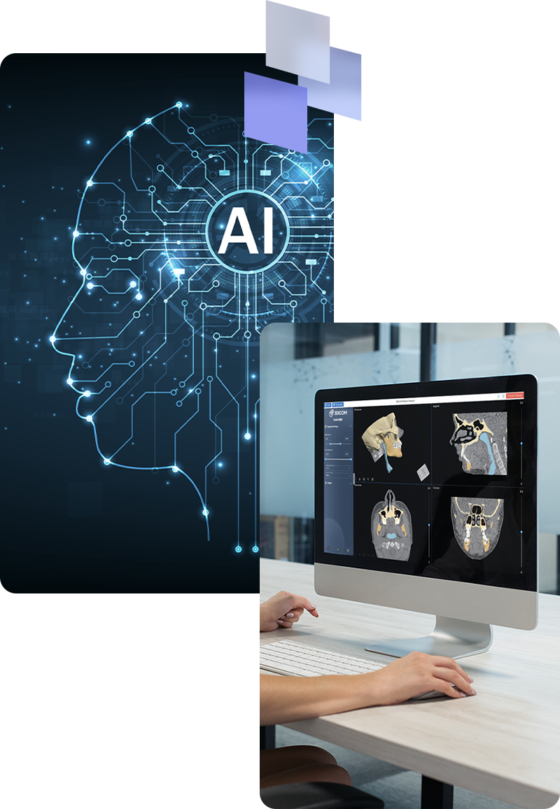
Expedite implant design R&D with AI-driven precision.
With 3DICOM EDU, you can harness patient DICOM data and a powerful library of Generative AI models like Relu to create patient-specific implants. Medical researchers and academics can use AI to automatically identify and populate model areas, generating custom implant solutions that can be easily exported for further R&D and prototyping.
Streamline R&D workflow with automated segmentation.
With access to automatic AI 3D model segmentation in 3DICOM EDU, specialists can quickly isolate anatomical structures, to save time and enhance accuracy, to create more time for deeper analysis. And efficiently create precise 3D models, essential for personalized treatment, research, and medical education.
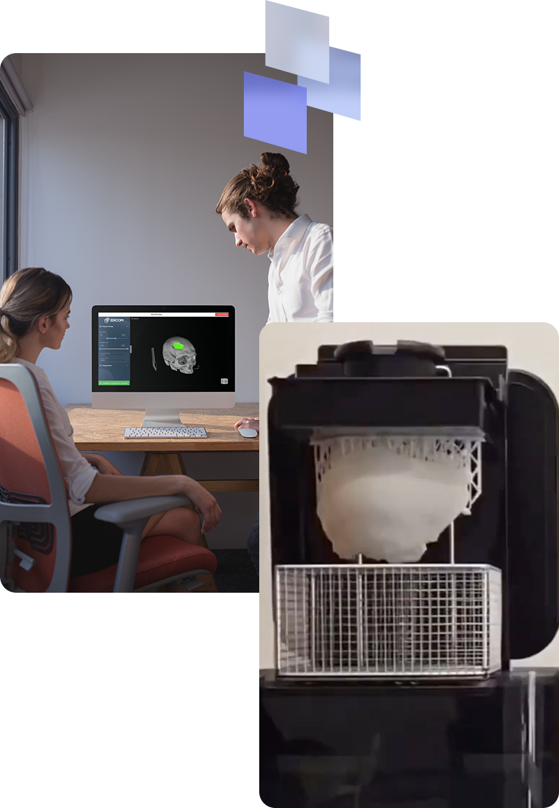
Create design files ready for 3D printing, in a snap!
Exporting design files for 3D printing from 3DICOM EDU is simple and efficient. With just a few clicks, 3DICOM EDU users can convert segmented patient-specific models and anatomical structures into print-ready .STL and .OBJ design files. This streamlined process saves time and ensures precision, making 3D printing accessible for medical research, education and prototyping.
Frequently Asked Questions
You’ll find answers to some of our most common questions below, to help you understand how 3DICOM EDU can fit into your current workflows.
How can 3DICOM EDU enhance our medical curriculum?
3DICOM EDU transforms 2D radiological images into interactive 3D models, allowing students to explore a wide range of anatomical structures, and pathologies in detail. This enhances understanding and engagement, making learning more effective and enjoyable. Additionally, with the optional inclusion of XR, the experience can become fully immersive for students.
What types of data can be imported into 3DICOM EDU?
3DICOM EDU supports a diverse range of data formats, including standard DICOM files for medical imaging and 3D MCAD design files in .OBJ and .STL formats. This versatility facilitates the importation of patient-specific imaging and integration of 3D-rendered medical devices, plates and screws, etc. Offering limitless possibilities for customized medical solutions.
Can 3DICOM EDU export models in formats compatible with most 3D printers?
Yes, 3DICOM EDU can export models in universally accepted formats such as STL and OBJ, which are compatible with a wide range of 3D printers. This flexibility allows for seamless transition from digital modeling to physical printing.
Can 3DICOM EDU be used for collaborative research projects?
Yes, the software offers advanced collaboration tools that enable teams to share and annotate 3D models, integrate clinical notes, and work synchronously, regardless of geographical locations.
Is there a steep learning curve associated with 3DICOM EDU?
3DICOM EDU is designed with a user-friendly interface to ensure ease of use for professionals across disciplines, as well as students. Additionally, there are comprehensive support and training materials to assist new users. These can be located in the Knowledge Base.
Can 3DICOM EDU assist in creating physical models through 3D printing?
Absolutely. The software can seamlessly export models to 3D printing formats, facilitating the creation of physical replicas for exploratory surgical planning, educational purposes, and research. Additionally, you’ll find a growing number of AI models accessible through the software, which expedites workflows for 3D printing patient-specific anatomical models and prosthetic devices.
Can 3DICOM EDU be used for educational purposes?
Yes, 3DICOM EDU excels as an educational tool, providing comprehensive 3D visualization, measurement, annotation, and markup capabilities. All annotations and markups can be fully viewed in 3D within the software and are included when sharing image sets in both 2D and 3D formats. This feature significantly enhances the ability of students, younger practitioners, nurses, and support staff to grasp the location and size of specific anatomical structures and pathologies securely and intuitively.

Unlock the full value of your medical imaging
Sign up today or try the FREE demo to experience the difference for yourself.
- For a full list of software inclusions and a comparison of features across 3DICOM products, please refer to the Pricing page. ↩︎