
Products
3DICOM Patient
View, understand, manage and share your medical images online or on the go with 3D visualization and AI education tools in 3DICOM Patient’s DICOM image viewer online.
SEE
what’s happening in your body!
Visualize your scans in 3D and explore your anatomy with AI for a clearer understanding. Empowering you to discuss your health confidently with your doctor and make informed decisions.
STORE
your scans in your pocket!
Say goodbye to physical records and CDs with instant access to your medical imaging wherever you are, whenever you need it, with 3DICOM’s online viewer and companion mobile app.
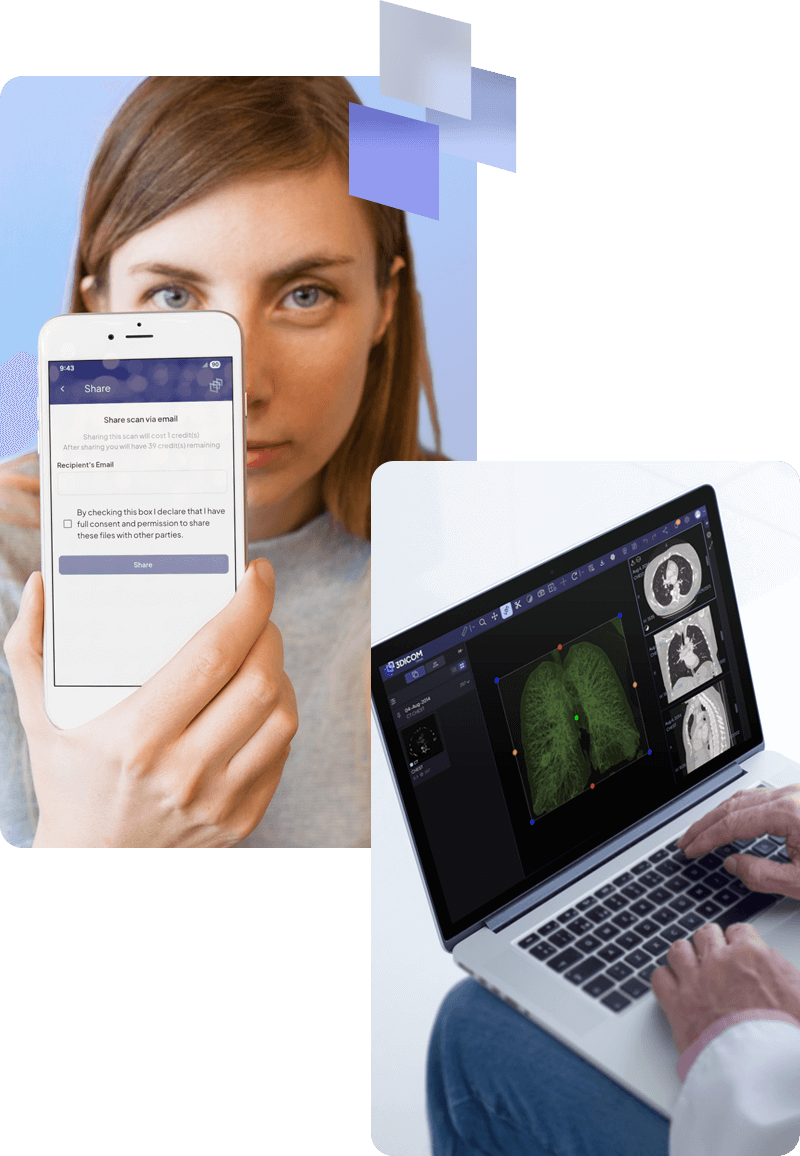
SHARE
your imaging fast, securely!
Avoid unnecessary duplicate imaging, saving time, money, and radiation exposure by quickly and securely sharing your scans with your care team whenever needed, with just one click.

What’s Included
3DICOM Patient is trusted by the following partners:
Enhance Understanding
SEE
what’s happening in your body with 3DICOM Patient!
See, learn, understand, and feel more empowered to make confident, well-informed decisions about your health using 3DICOM Patient.
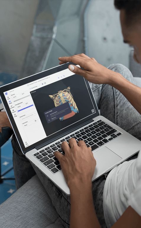
Transform 2D medical images into 3D, effortlessly!
With just a click, 3DICOM Patient transforms complex 2D images from CT, MRI, and PET scans into interactive 3D models, that you can rotate, pan and zoom to see from every angle.
Use AI to explore and learn about your body’s anatomy.
With the assistance of artificial intelligence, which segments and labels areas of your personalized 3D model, you can easily identify the parts your doctor is discussing, to further enhance your understanding of your medical condition.
Communicate more effectively with your doctor.
By using 3DICOM Patient’s advanced visualizations and AI integrations, you can better understand your body, enhance discussions with your doctor, and feel confident in making well-informed decisions about your health.
Convenient Storage
STORE
your scans in your pocket with 3DICOM Patient!
Simplify your medical image storage like never before with 3DICOM Patient.
Say goodbye to physical records and outdated CDs.
With the ability to store your medical imaging on any internet-capable device (computer, phone, or tablet) using 3DICOM Patient you can finally #DitchtheDisk.
Store your medical images securely and long-term.
While an internet connection is required to log in and receive updates, your medical data (DICOM files) is stored and processed locally on your device, ensuring that your personal information stays secure.
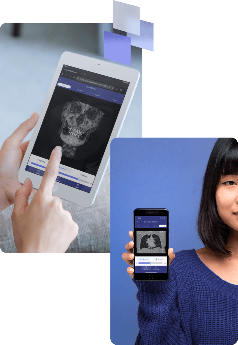
Access your medical imaging anytime.
Keep your medical imaging securely stored on any device, ensuring convenient, instant access anytime you need it, no matter where you are.
Fast & Secure Sharing
SHARE
your imaging fast, securely with 3DICOM Patient!
Embrace a simple, secure way to share your imaging records fast using 3DICOM Patient!
Share your imaging with one click, anytime and from anywhere.
With the 3DICOM Patient mobile app or online viewer, sharing your medical images with your doctor or specialist is as simple as hitting ‘send,’ streamlining your care and enhancing your connection to your healthcare team.
Keep control of your imaging records.
With 3DICOM Patient’s HIPAA-compliant solution, designed to protect your privacy through advanced encryption and robust security, you can rest assured your medical images and data stay safe during transfers, letting you focus on your health without privacy concerns.
Save time and money on your care with 3DICOM Patient.
By having your medical images readily available to share with healthcare providers, you can avoid unnecessary duplicate imaging, helping you prevent treatment delays, extra imaging costs, and unnecessary radiation exposure.
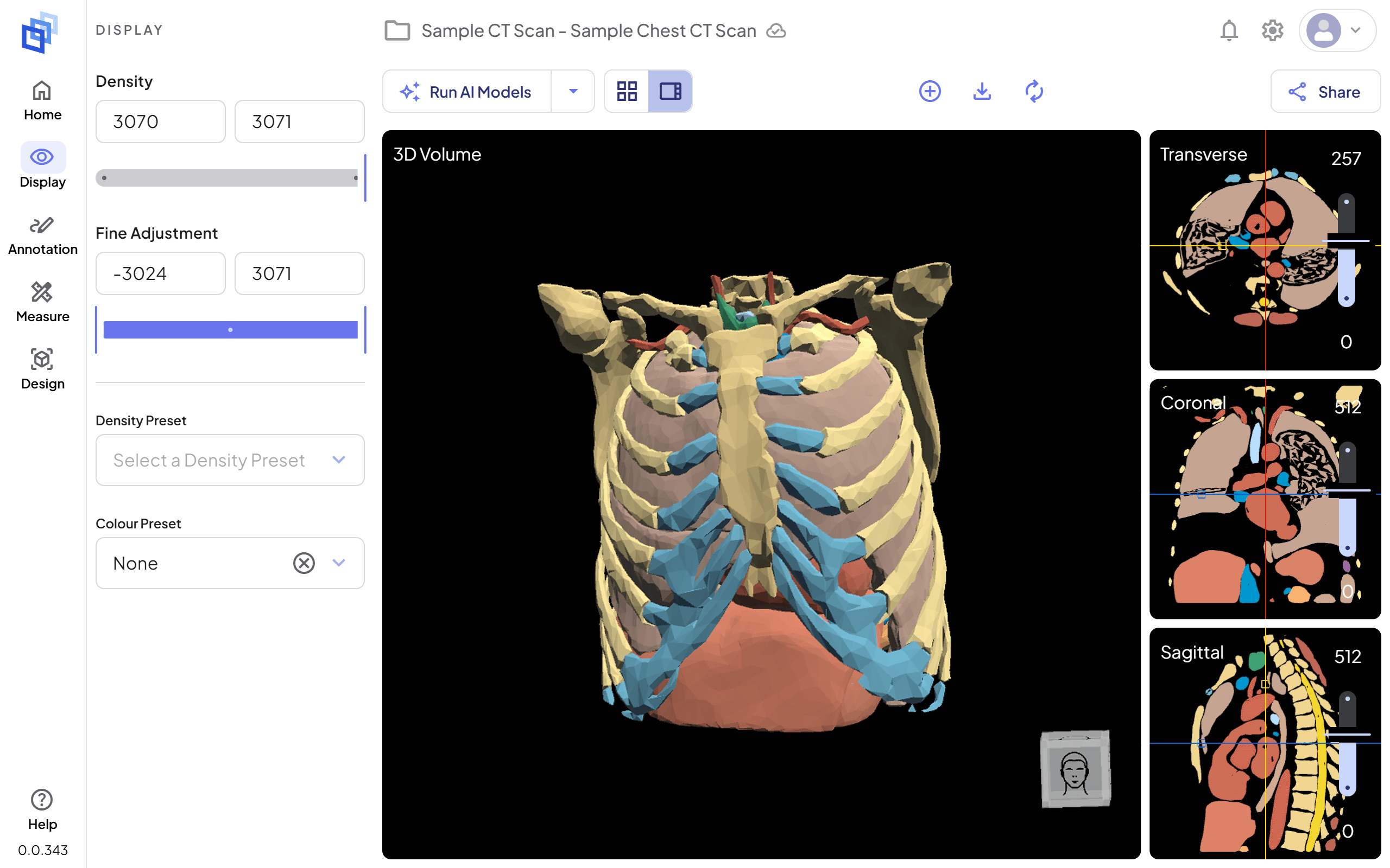
DICOM Viewer Example Screenshot – 3DICOM Patient
How can I view DICOM images on my computer?
The 3DICOM Patient online viewer lets you view your DICOM images online on your computer using any internet browser. All you need is a stable internet connection. Subscribe to access the 3DICOM Patient viewer, upload your first DICOM images and follow the built-in tutorial to view them in 3D to make them easier to understand.
Frequently Asked Questions
Here are answers to some of our most common questions to ensure you feel empowered to access, view, store, and share your medical imaging.
What is 3DICOM Patient software?
3DICOM Patient is an advanced medical imaging viewer that transforms traditional 2D scans like MRI, CT, and PET into interactive 3D models. It equips patients with intuitive tools to explore and understand their medical data, enhancing visualization and personal health insights. Empowering patients further, it offers full control to access, store, and share images across devices and with healthcare professionals anytime, anywhere.
What is a DICOM file?
DICOM (Digital Imaging and Communications in Medicine) is a worldwide standard file format for storing, sharing, and viewing medical images, such as MRI, CT, and PET scans. This format ensures that images can be accessed and read on different devices using specialized software called DICOM viewers, like 3DICOM Patient, regardless of the imaging machine, body part, or location where the image was taken.
How can I view DICOM images on my computer?
The 3DICOM Patient online viewer lets you view your DICOM images online on your computer using any internet browser. All you need is a stable internet connection. Subscribe to access the 3DICOM Patient viewer, upload your first DICOM images and follow the built-in tutorial to view them in 3D to make them easier to understand.
Is 3DICOM Patient software easy to use for someone without a medical background?
Yes, the software is designed with a user-friendly interface that makes it accessible for individuals without medical training. When first using the software, you will be introduced to the functionalities within the online dashboard and shown how to navigate the viewer through a guided tutorial when you open your first scan.
Additionally, as part of our commitment to providing the best possible user experience, we greatly appreciate your feedback. Your input is crucial in helping us continually enhance the software for all users. Should you encounter any difficulties, you can submit your request for support through the 3DICOM online viewer.
Do I need any special equipment to use 3DICOM Patient software on my computer?
No special equipment is needed. You can access the 3DICOM Patient online viewer from any internet-enabled computer or tablet. However, it is recommended to use a device with good screen resolution to better view the your imaging. Additionally, you can also securely share your medical imaging to your mobile device for convenient access, using the 3DICOM Mobile app.
How do I obtain my medical images to use in the 3DICOM Patient software?
You can obtain your medical images from your healthcare provider, who can give you a copy on a CD, DVD, or a secure digital download link. These images are typically in the DICOM format, which you can open in 3DICOM Patient software.
What do I need to start viewing my body in 3D using 3DICOM Patient?
To get started, all you need is the DICOM file from your hospital or imaging provider and an internet-enabled device (computer, phone, or tablet). 3DICOM Patient works across platforms, so it’s compatible with Windows, Mac, iOS, and Android devices.
Can I share my images from 3DICOM Patient with another doctor?
Yes, 3DICOM software allows you to securely share your images. You can send your medical imaging files directly to your care provider through the platform, making it easy to consult with other specialists if needed. All you need is your doctor’s email address.
Can I use the software to measure dimensions of a tumor or other medical features?
While this is not possible in 3DICOM Patient to take measurements, medical practitioners using 3DICOM MD have access to a number of tools that allow them to measure distances, areas, and other features within your medical images, which can then be shared with you. This can be useful for tracking changes over time. And generally improving your understanding of what is occurring in your body.
Does 3DICOM Patient provide any educational content to help me understand my scans?
Yes, 3DICOM Patient includes a variety of educational tool tips and integrations within the software to help you navigate the interactive DICOM viewer, annotate, and learn about different areas of the body, enhancing your understanding of what you are viewing.
Additionally, you can access a wealth of educational resources, under the Resource section of the 3DICOM website.
What should I do if I have trouble using the software?
3DICOM’s extensive Knowledge Base offers a wealth of online resources and training materials, providing helpful tips and guidance for making the most of the software.
Should you require further assistance, or encounter any technical issues with the software our dedicated 3DICOM support staff are readily available to assist you via email. Support tickets can be lodged via the viewer dashboard.
Can I view images from my smartphone or tablet using 3DICOM Patient?
Yes, there is a mobile version or web-based version of the software that is compatible with smartphones and tablets, making it convenient to view your medical images on the go.
Is 3DICOM Patient a free DICOM reader or viewer?
While the demo version of 3DCIOM Patient is entirely free to access, it is not possible to upload your own medical scans for viewing in this version. To unlock the full version of 3DICOM Patient, including all features and functionality, a subscription is required. Flexible options are available with affordable monthly or annual plans. For more information, please visit the 3DICOM Pricing and Features page.

Unlock the full value of your medical imaging
Sign up today or try the FREE demo to experience the difference for yourself.
Learn more about our story
Read about 3DICOM’s beginnings and mission on our about page.