The Benefits of 3D Viewing Scans of Cranial and Spinal Foramina
Cranial and Spinal Foramina send important signals to the body and play a large part in our overall health. There are a range of benefits in 3D Viewing scans of Cranial and Spinal Foramina, which are listed below.

In the realm of medical imaging, particularly in evaluating cranial and spinal foramina, advancements in technology have dramatically changed how radiologists and healthcare professionals assess and interpret scans.
Understanding Cranial and Spinal Foramina
Cranial and spinal foramina are critical structures within the human body that play essential roles in nerve function and overall health. These passageways allow for the safe exit of nerves and blood vessels from the skull and the vertebral column to other parts of the body.
Each foramina’s anatomy is distinct, with specific characteristics that define its functions. Understanding these structures is crucial for diagnosing and treating various medical conditions.
Anatomy of Cranial and Spinal Foramina
The cranial foramina include several openings in the skull, such as the foramen magnum, where the spinal cord passes through and connects to the brain. Other notable foramina include the optic canal, which allows the optic nerve to enter the orbit, and the jugular foramen, permitting vital vessels and nerves to exit the skull.
Similarly, the spinal foramina are openings in the vertebrae that allow for the escape of spinal nerves. Each spinal segment has its unique foramina that corresponds to specific nerves. This complex anatomy necessitates precise imaging techniques to visualize any obstructions or anomalies effectively. The foramina’s sizes and shapes can vary significantly among individuals, which can influence the risk of nerve compression or injury, making personalized medical assessments a necessity.
Common Disorders and Conditions
Several disorders can affect cranial and spinal foramina, including herniated discs, spinal stenosis, and tumors. Conditions such as Chiari malformation can impact the functioning of the cranial foramina, leading to significant neurological symptoms. These disorders highlight the importance of accurate imaging to facilitate timely diagnosis and management.
Moreover, trauma can lead to fractures affecting these foramina, putting nerves at risk and necessitating surgical intervention. Understanding these conditions further emphasizes the need for advanced imaging capabilities like 3D viewing.
In addition to trauma, degenerative diseases such as osteoarthritis can contribute to the narrowing of foramina, a condition known as foraminal stenosis, which can cause chronic pain and mobility issues. Patients experiencing symptoms such as numbness, tingling, or weakness in the limbs should be evaluated for potential foraminal involvement, as early intervention can significantly improve outcomes.
The Rise of 3D Imaging
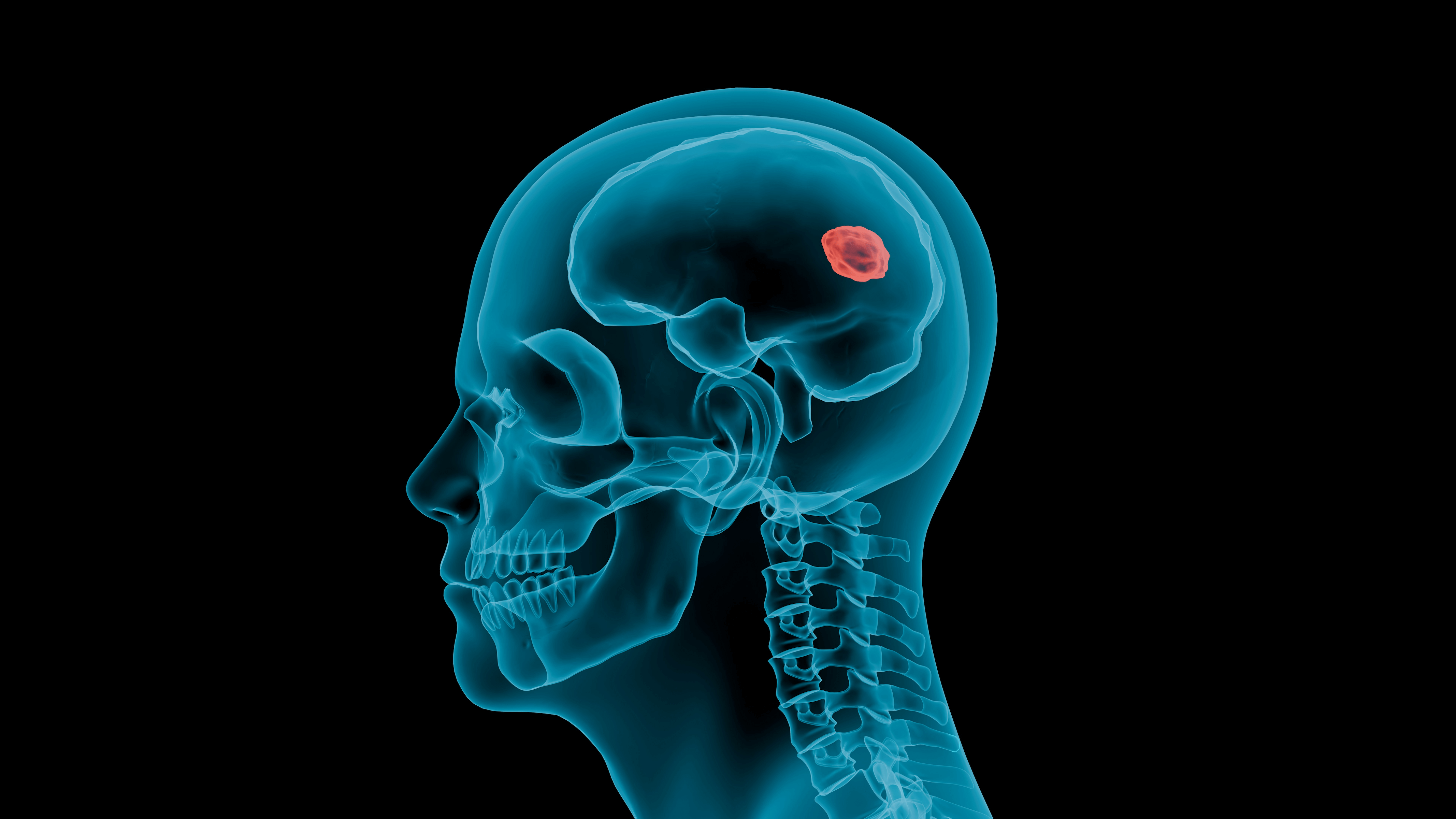
The introduction of 3D imaging represents a significant step forward in medical diagnostics. By generating 3D reconstructions from 2D scan data, healthcare providers can visualize structures in a more intuitive and detailed way. 3DICOM MD is one of the 3D Viewing platforms ideal for physicians.
This advancement allows for better assessment of foramina and surrounding tissues, leading to enhanced diagnostic accuracy. The capability to virtually manipulate images provides insights that traditional methods cannot offer.
Additionally, 3D imaging has revolutionized surgical planning, enabling surgeons to rehearse complex procedures using accurate anatomical models before entering the operating room. This not only improves surgical outcomes but also reduces the risk of complications, as physicians can anticipate challenges based on precise anatomical visualizations. The integration of AI in 3D imaging further refines diagnostics, offering predictive analytics and automated anomaly detection that can significantly enhance patient care.
Improved Diagnosis and Treatment Planning
With improved imaging capabilities comes better diagnosis. The detailed views provided by 3D scans allow for a more accurate assessment of various conditions affecting the cranial and spinal foramina. By understanding the complexities of these structures, healthcare providers can formulate targeted treatment strategies.


