2D and 3D Diagnosis & Planning with
3DICOM for Veterinary Medicine
Disclaimer: 3Dicom R&D is NOT a Medical Device and is intended for research, scientific & educational purposes only.
3D volume-render, manipulate, measure and annotate veterinary images at the convenience of your own device

Complete your understanding of veterinary imaging and gain additional radiological information with 3Dicom R&D’s intuitive visualisation features.
In a matter of seconds, watch how conventional veterinary scans transform into fully-immersive 3D models, where you can zoom in on, pan and rotate the 3D body of an animal, isolate particular regions of interest and use the 3D intersect tool to gain precise knowledge of the case at hand.
Furthermore, the in-built Housfield Unit Window feature allows users to distinguish between an animal’s skin, tissue and bones. You can also view veterinary scans with low opacity as an X-RAY type image or increase the opacity to further define key areas of interest.
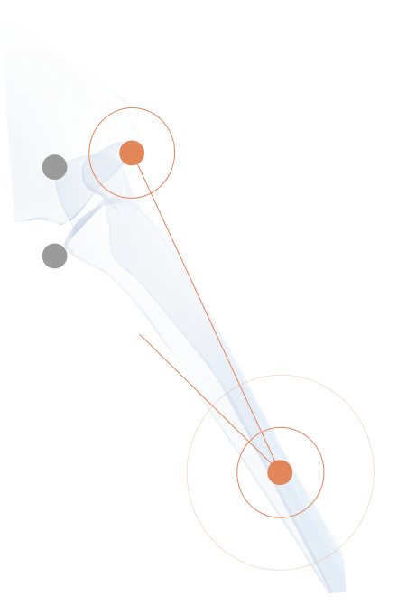
Veterinary doctors, nurses and even students will also find 3Dicom R&D’s annotation and measurement tool useful to gain an insight on the distances, areas and even angles which can be determined directly from the 2D scan uploaded into 3Dicom. These annotations and measurements can then be viewed in 3D and securely shared between the team.
Test veterinary implants and plan surgical intervention with 3Dicom R&D’s MCAD feature
Whether you need to test the fit of an implant for a canine recieving a joint replacement, or manage neck and vertebral fractures in a horse, you can do so using 3Dicom R&D’s MCAD feature.
This feature allows you to easily import MCAD files into the 3Dicom software, simulate specific surgical intervention of bespoke implants and for added precision, explore exactly how the implant will sit inside the animal’s actual body.

Convert 2D veterinary CT and MRI images into 3D printable models within minutes
Veterinary doctors wanting an intuitive platform to 3D print bespoke implants for complicated case situations can now do so with 3Dicom R&D’s complete set of segmentation tools now available with different colours and labels.
The segment to 3D print process can also be used for educational purposes to better inform veterinary team members of the specific region of interest. The process is completed using semi-automated techniques such as threshold flood-fill, level tracing and island removal with small edits made manually.
The segmented anatomy can then be exported to STL, OBJ or PLY file types for use in additive manufacturing or for the printing of physical 3D anatomical models.
Enhance the knowledge and understanding of veterinary cases to your team and pet-owners
3Dicom R&D was designed with the purpose to educate and help those in the veterinary field gain a more precise understanding of the case at hand. With detailed visualisation capabilities such as the option to colour render veterinary images, you can gain added context on the spacial relationship of anatomy within an animal’s body.
3Dicom R&D is a worthwhile addition to your workspace as veterinary doctors, nurses and even animal owners can visually comprehend and interact with the area of interest.