The Purpose and Benefits of Musculoskeletal 3D Imaging
Explore the transformative impact of musculoskeletal 3D imaging in modern healthcare.

In recent years, advancements in medical imaging technology have revolutionized the way healthcare professionals diagnose and treat musculoskeletal conditions.
One of the most significant developments in this field is musculoskeletal 3D imaging. This technology offers a detailed view of bones, muscles, and joints, providing invaluable insights that traditional imaging methods cannot match. Understanding the purpose and benefits of musculoskeletal 3D imaging can help patients and healthcare providers make informed decisions about diagnosis and treatment options.
Understanding Musculoskeletal 3D Imaging
What is Musculoskeletal 3D Imaging?
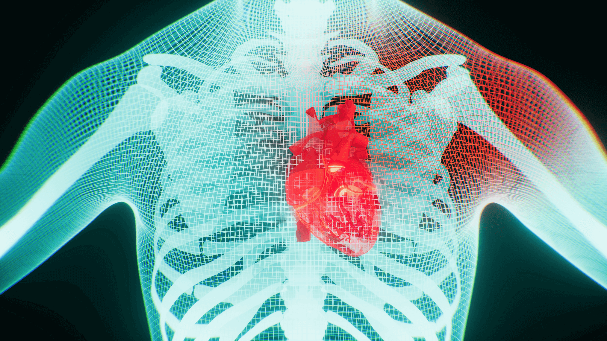
Musculoskeletal 3D imaging is a sophisticated imaging technique that creates three-dimensional representations of the body’s musculoskeletal system. Unlike traditional X-rays or 2D scans, 3D imaging provides a comprehensive view of the anatomical structures, allowing for a more accurate assessment of injuries, diseases, and abnormalities. This technology utilizes advanced software, such as 3DICOM, and imaging equipment to capture detailed images that can be rotated and viewed from multiple angles.
3D imaging is particularly useful in visualizing complex structures such as joints, where overlapping bones and tissues can obscure critical details in 2D images. By offering a more complete picture, 3D imaging aids in precise diagnosis and treatment planning, ultimately improving patient outcomes.
How Does Musculoskeletal 3D Imaging Work?
The process of musculoskeletal 3D imaging involves several steps. Initially, a series of 2D images are captured using advanced imaging modalities such as CT or MRI. These images are then processed using 3DICOM or other specialized software, that reconstructs them into a 3D model. The resulting model can be manipulated to view different angles and cross-sections, providing a comprehensive understanding of the area of interest.
Healthcare providers can use these 3D models to identify subtle changes in bone density, detect fractures, assess joint alignment, and evaluate soft tissue conditions. This level of detail is particularly beneficial in complex cases where traditional imaging may fall short.
The Purpose of Musculoskeletal 3D Imaging
Enhanced Diagnostic Accuracy
One of the primary purposes of musculoskeletal 3D imaging is to enhance diagnostic accuracy. By providing a detailed view of the musculoskeletal system, healthcare providers can identify conditions that may not be visible on standard 2D images. This is especially important in cases involving small fractures, ligament tears, or early-stage arthritis, where early detection can significantly impact treatment outcomes.
Accurate diagnosis is crucial for developing effective treatment plans. With 3D imaging, doctors can pinpoint the exact location and extent of an injury or disease, enabling them to tailor interventions to the patient’s specific needs.
Pre-Surgical Planning and Assessment
Musculoskeletal 3D imaging plays a vital role in pre-surgical planning and assessment. Surgeons can use 3D models to visualize the surgical site, plan the procedure, and anticipate potential challenges. This level of preparation reduces the risk of complications and improves surgical precision, leading to better patient outcomes.
In addition to aiding in surgical planning, 3D imaging allows for post-operative assessment. Surgeons can evaluate the success of the procedure by comparing pre- and post-surgery images, ensuring that the desired outcomes have been achieved.
The Benefits of Musculoskeletal 3D Imaging
Improved Patient Experience
Musculoskeletal 3D imaging offers several benefits that enhance the overall patient experience. One of the most notable advantages is the reduction in the need for exploratory surgery. By providing a clear view of the affected area, 3D imaging can eliminate the need for invasive diagnostic procedures, reducing patient discomfort and recovery time.
Additionally, the detailed images produced by 3D imaging can help patients better understand their condition. By visualizing their anatomy, patients can gain a clearer understanding of their diagnosis and treatment options, leading to more informed decision-making and increased satisfaction with their care.
Facilitating Research and Innovation
Musculoskeletal 3D imaging is not only beneficial for clinical practice but also plays a crucial role in research and innovation. Researchers can use 3D models to study the biomechanics of the musculoskeletal system, leading to new insights and advancements in treatment options. This technology also supports the development of personalized medicine, where treatments are tailored to the individual characteristics of each patient.
Musculoskeletal 3D imaging represents a significant advancement in medical imaging, offering numerous benefits for both patients and healthcare providers. By providing detailed and accurate images of the musculoskeletal system, technology like 3DICOM, enhances diagnostic accuracy, improves surgical planning, and facilitates research and innovation.
As musculoskeletal 3D imaging continues to evolve, it holds the promise of transforming the way musculoskeletal conditions are diagnosed and treated, ultimately leading to better patient outcomes and improved quality of care.


